
When the Pelvis Stops Guiding the Spine
New European Spine Journal research — and why it lets us give more precise care at Symmetry Health Center
Quality care is not a slogan. It is a measurement you can defend. A new paper in the European Spine Journal (a Q1 orthopedic spine journal) gives our clinic a clearer way to do that for people with chronic low back pain: stop treating a generic lumbar curve, and start treating the curve that matches that patient’s own pelvic shape.
That is how this research shows up in the treatment room. It does not replace listening to the patient. It gives us a radiographic target so the plan is individualized instead of average.
Read the study (open access PDF):
https://link.springer.com/content/pdf/10.1007/s00586-026-10374-9.pdf
Harrison DE, Colloca CJ, Oakley PA, Moustafa IM. Pelvic morphology decoupling from lumbar lordosis in chronic low back pain. Eur Spine J. 2026. doi:10.1007/s00586-026-10374-9
This is the 16th CBP NonProfit publication of the year. A grant from NCMIC to CBP NonProfit partially funded the project. Independent research like this is what allows us to give quality care — care grounded in published biomechanics, not guesswork.
The study compared 72 people with chronic low back pain (CLBP) to 72 asymptomatic volunteers using standing lateral full-spine radiographs.
In healthy spines, morphology still drives the curve
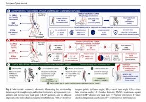
In asymptomatic volunteers, pelvic shape (PTPIA — a fixed anatomical constant) and sacral base angle (SBA) work together to constrain lumbar lordosis (L1–L5 ARA).
- PTPIA and lordosis were significantly correlated (r = –0.428, p < 0.001).
- A multivariate model (PTPIA + SBA + T3–T10 kyphosis) explained about 32.5% of lordosis variance.
- Prediction error was lower (RMSE ≈ 9.4°).
That is an intact coupling chain: pelvic morphology → sacral inclination → lumbar curve.
In chronic low back pain, that chain breaks
In the CLBP group the PTPIA–lordosis link disappeared (r = –0.137, p = 0.251). SBA became the dominant driver, with a roughly three-fold amplified coefficient (β ≈ –1.32 vs –0.40 in normals).
Radiographs in that group also showed the posture of pelvic retroversion:
- lower SBA
- higher pelvic tilt
- greater S1-to-hip-axis distance
Mean L1–L5 lordosis was not dramatically different between groups. The problem was mismatch, not a single average number.
About 44.4% of CLBP patients had less lordosis than their own pelvic morphology predicted (positive “lordosis gap,” mean deficit ≈ +3.87°). The rest sat on the other side of the equation (more curve than expected).
Why this lets us give better care
A “normal-looking” lumbar angle is not enough. Quality care asks a better question:
Does this patient’s lordosis match their pelvic morphology?
If it does not, we now have a published, step-by-step way to set the target:
- Measure PTPIA on a standing lateral radiograph.
- Calculate expected lordosis from the normal-group relationship
(Expected LL = –0.312 × PTPIA_adj + 14.96). - Measure actual L1–L5 lordosis (posterior tangent / ARA).
- Compute the lordosis gap (actual minus expected).
- Positive gap (hypolordosis / under-curved) → increase lordosis.
- Negative gap (hyperlordosis / over-curved) → decrease lordosis.
That gap is what we treat. Not a textbook number. Not “strengthen your core and hope.” A morphologically informed sagittal target for that patient.
How this looks at Symmetry Health Center
We use standing full-spine radiography and CBP®-style sagittal analysis so rehabilitation matches the film, not a protocol sheet.
Depending on the gap, care may include:
- extension-based and elongation work (including Schroth / ScoliBalance-style “grow tall” cues when deformity is also present)
- pelvic nutation / anti-retroversion positioning
- mirror-image exercise and traction when films support it
- progress tracked on repeat standing radiographs, not only pain scores
The paper is cross-sectional, so it cannot prove that retroversion causes pain. Age and sex differed between groups; the authors’ sensitivity analyses still held. What it gives our patients is something more practical: a defensible outcome measure. We can show whether the curve is moving toward the value their own pelvis predicts.
That is quality care — measurable, individualized, and honest about what the X-ray actually shows.
If you have been told your X-rays are “fine”
Many people with months of low back pain hear that films are unremarkable because no fracture, listhesis, or “surgical” curve is present. This study says the missing piece is often decoupling — the lumbar spine has stopped tracking pelvic shape, and sacral inclination is running the show.
That is measurable. It is also treatable as a target, not a mystery.
Support the research behind this kind of care
Work like this does not happen in a vacuum. CBP NonProfit funds and publishes studies that test whether spinal alignment actually matters — and how to measure it well enough to treat it. If patients deserve care built on evidence instead of habit, that research has to keep getting done.
Support CBP NonProfit: cbpnonprofit.com
A portion of this project was supported by an NCMIC grant to CBP NonProfit. Thank you to the research team: Deed Harrison, Chris Colloca, Paul Oakley, and Ibrahim Moustafa.
Next step
If you have chronic low back pain and have not had a standing full-spine analysis that includes pelvic morphology and a lordosis-gap calculation, that is the visit to schedule. Bring prior films if you have them. We will tell you whether your curve still matches your pelvis — and build a plan around that answer.
Symmetry Health Center / Bay Area Scoliosis Center
1708 Park St. Suite 130, Alameda, CA 94501
Reference
Harrison DE, Colloca CJ, Oakley PA, Moustafa IM. Pelvic morphology decoupling from lumbar lordosis in chronic low back pain. European Spine Journal. 2026. https://doi.org/10.1007/s00586-026-10374-9
Direct PDF: https://link.springer.com/content/pdf/10.1007/s00586-026-10374-9.pdf
CBP NonProfit: https://cbpnonprofit.com